
Expert Endometriosis Ultrasound — ISUOG IDEA Consensus Protocol
Expert transvaginal ultrasound is the first-line investigation for endometriosis. Performed using the ISUOG IDEA consensus protocol by an experienced sonographer, it detects most deep infiltrating disease without surgery — fundamentally changing how endometriosis is diagnosed in 2026.
1. What IDEA stands for
IDEA — International Deep Endometriosis Analysis — is an ISUOG (International Society of Ultrasound in Obstetrics and Gynecology) consensus statement published in 2016 and updated in 2024. It standardises systematic ultrasound examination for endometriosis, ensuring consistent, reproducible mapping of disease across all pelvic compartments.
2. The four compartments examined
Anterior compartment: bladder, uterovesical fold, anterior uterine wall. Posterior compartment: uterosacral ligaments, torus uterinus, rectovaginal septum, anterior rectum and rectosigmoid. Ovaries: endometriomas, mobility, kissing-ovaries sign, ovarian reserve markers. Uterus: junctional zone, adenomyosis features, myometrial cysts, fibroids.
3. Soft markers and dynamic assessment
Beyond static lesion identification, IDEA includes dynamic ultrasound assessment: ovarian mobility (fixed ovary suggests adhesion), sliding sign (uterus and rectum slide independently when not tethered), bladder distensibility, peristalsis of bowel near lesions. These dynamic findings provide critical surgical-planning information.
4. What ultrasound detects accurately
Endometriomas (95+% sensitivity in expert hands). Deep infiltrating lesions of uterosacrals, rectovaginal septum, posterior fornix (85-90% sensitivity). Bladder DIE (90%+ sensitivity). Bowel DIE (80-85% sensitivity, with depth and distance measurement). Adenomyosis (MUSA criteria, 80-90% sensitivity). Pelvic adhesions (via dynamic sliding sign).
5. What ultrasound may miss
Superficial peritoneal endometriosis (lesions less than 5 mm on peritoneal surface) often not detectable on ultrasound. Ureteric endometriosis may need additional MRI urography. Diaphragmatic or thoracic endometriosis requires extended imaging. Very small lesions deep in the bowel wall may be ambiguous.
6. Sonographer expertise matters
The IDEA protocol requires specific training and experience. A general radiographer doing a routine pelvic ultrasound is not performing the IDEA protocol. Detection rates differ enormously between expert and non-expert ultrasound — this is the main reason endometriosis is missed on standard scanning.
7. What to expect during the scan
Transvaginal ultrasound (more sensitive than transabdominal for endometriosis). Lasts 30–45 minutes for full IDEA examination. Mildly uncomfortable but not painful. Empty bladder before examination. Findings discussed at end of scan with imaging shown. Written report provided.
8. Combining ultrasound with other investigations
Expert ultrasound is the first-line investigation. MRI adds detail for surgical planning of complex deep disease, bowel/bladder involvement, ureteric mapping. The two modalities are complementary, not alternatives. Both inform individualised treatment planning.
What an expert (IDEA-protocol) scan checks
| Region | What we assess |
|---|---|
| Ovaries | Endometriomas, mobility, “kissing ovaries” |
| Uterus | Adenomyosis features, mobility |
| Pouch of Douglas | Obliteration (sliding sign) |
| Bowel (rectum / sigmoid) | Deep nodules and their depth |
| Bladder & ureters | Nodules, dilatation |
Aligned with current international evidence, not habit.
Frequently Asked Questions
How accurate is expert ultrasound for endometriosis?
Is the scan painful?
How long does the scan take?
Should I prepare for the scan?
Can routine ultrasound diagnose endometriosis?
Will I need MRI as well?
What is the sliding sign?
What if my previous ultrasound was normal?
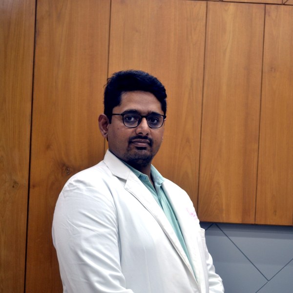
Dr Patel leads endometriosis diagnosis and surgery at Balaji Horizon with an evidence-based, ovarian-sparing philosophy aligned to ESHRE and ESGE — integrating pain, fertility and long-term disease control into a single plan, rather than treating the disease in isolation.
Imaging-led diagnosis, medical-first management, and precise surgery only when it is the right step — planned around your pain and fertility goals.
Fetal sex is neither determined nor disclosed, as per the PC-PNDT Act.
Dr. Priyadatt Patel
Senior Gynecologist · Advanced Laparoscopic Surgeon · IVF and Endometriosis Programme Lead
MS OBGyn · Pregnancy Care · Advanced Gynaecological Ultrasound · Fertility Preservation
ESHRE / ESGE / AAGL / ASRM guideline-aligned practice. 3D Karl Storz precision technique. Fertility-preservation-first philosophy. Evidence-based decisions, honest counselling, long-term outcomes orientation.
323, Satyamev Eminence, Beside Saptak Bungalows & AUDA Water Tank, Science City Road, Sola, Ahmedabad 380060
Open 24 hours (emergencies & labour) · OPD Mon–Sat 11:00–20:00 · +91 97234 31544
Naranpura, Ahmedabad
Mon–Sat 08:30–10:30 · +91 70460 02566