
Endometriosis · Complex disease
Deep infiltrating endometriosis — a reference for patients and referring clinicians
Deep infiltrating endometriosis (DIE) is endometriosis that has invaded beneath the peritoneal surface by more than 5 millimetres — the most clinically demanding form of the disease. At Balaji Horizon Women’s Hospital, the endometriosis programme is led by Dr. Priyadatt Patel — Senior Gynecologist, Advanced Laparoscopic Surgeon, and IVF & Endometriosis Programme Lead — with the Diploma in Gynaecological Endoscopy from the Kiel School of Gynaecological Endoscopy (Germany) and the Part 1 Diploma in Operative Endoscopy, ESGE-CICE (France). The programme has handled 500+ endometriosis cases and 3,000+ advanced laparoscopic procedures across 13+ years, and runs to ISUOG IDEA imaging standards. This page is written for patients seeking a clear understanding before consultation and for referring clinicians who want to know how the centre approaches DIE.
What is deep infiltrating endometriosis?
Endometriosis exists on a spectrum. Superficial peritoneal disease sits on the surface of the pelvic lining. Ovarian endometriomas are cystic. Deep infiltrating endometriosis penetrates more than 5 millimetres into tissue — uterosacral ligaments, the recto-vaginal septum, the bowel wall, the bladder wall, the ureters, or, less commonly, the pelvic side-wall and beyond.
Because DIE involves structures outside the standard gynaecological field, accurate mapping before surgery and a multidisciplinary surgical team are central to safe, effective treatment.
Why DIE matters clinically
DIE drives the most disabling symptoms of endometriosis — severe deep dyspareunia, chronic pelvic pain that does not respond to standard analgesia, cyclical bowel and bladder symptoms, and infertility. It is also where surgical complications are most likely if the surgery is performed without adequate planning. Recurrence after incomplete excision is well documented, and the cost of a re-do operation includes both the patient’s recovery burden and a measurable impact on ovarian reserve where the ovaries are involved.
For these reasons, DIE is the form of endometriosis that most often warrants referral to a centre with experience in advanced excision.
Common patterns of involvement
- Uterosacral ligaments — commonly the first deep site; nodular thickening palpable on examination
- Recto-vaginal septum and posterior cul-de-sac — obliterates the pouch of Douglas; often presents with deep dyspareunia and dyschezia
- Bowel wall (rectum, sigmoid, less often ileum or appendix) — cyclical change in bowel habit, dyschezia, occasionally cyclical bleeding
- Bladder and ureters — dysuria, urgency, occasional cyclical haematuria; silent ureteric obstruction is the dangerous variant
- Pelvic side-wall — can involve nerves and major vessels; planning is critical
Multiple sites are common; isolated single-site DIE is the exception, not the rule.
Symptoms that suggest DIE
- Severe, cyclical, or chronic non-cyclical pelvic pain
- Deep dyspareunia — pain on deep penetration, often persisting after intercourse
- Dyschezia — painful bowel motions, often cyclical
- Cyclical haematuria, dysuria, or urinary urgency
- Cyclical haemoptysis or shoulder-tip pain (rare; suggests thoracic or diaphragmatic involvement)
- Subfertility, often alongside other features
- Symptoms that do not respond to standard hormonal therapy
Many patients carry these symptoms for years before the link to endometriosis is made. The pattern is the diagnostic clue.
Diagnostic approach
DIE is diagnosed clinically, with imaging confirmation and surgical correlation. The work-up at this centre follows a structured pathway:
- Structured history — chronology of symptoms, cycle pattern, sexual and bowel-bladder review, fertility goals, previous surgery
- Pelvic examination — targeted to detect nodularity of the uterosacrals, tender fixed adnexae, recto-vaginal nodules
- Transvaginal ultrasound (TVS) following the ISUOG IDEA protocol — a structured, systematic scan that reports each anatomical compartment
- MRI of the pelvis — for surgical mapping, particularly where bowel, bladder, ureter, or side-wall disease is suspected
- Additional studies where indicated — renal ultrasound and split renal function if ureteric involvement is suspected; cystoscopy for bladder disease; colonoscopy for differential diagnosis where bowel symptoms dominate
For further detail on the imaging pathway, see imaging for endometriosis.
All diagnostic ultrasound at Balaji Horizon is performed following the ISUOG IDEA (International Deep Endometriosis Analysis) consensus protocol — Dr. Patel is formally trained in this systematic four-compartment approach, which substantially out-performs generic pelvic ultrasound for detecting deep infiltrating disease. For surgical mapping cases, MRI is reviewed jointly by the operating team before the consultation closes.
Treatment principles
Treatment is individualised. The decision rests on symptom burden, ovarian reserve, fertility goals, organ involvement, age, and the patient’s priorities. Three broad pathways exist:
- Conservative medical therapy — progestogens, combined hormonal contraception, GnRH analogues with add-back, dienogest. Useful for symptom control where surgery is not yet indicated or while planning fertility treatment.
- Excisional surgery — complete removal of deep nodules, peritoneal disease, and any endometriomas, with restoration of anatomy. Excision (not ablation) is preferred for DIE per current ESHRE and ESGE guidance.
- Integrated IVF planning — in patients trying to conceive, the sequence of surgery and IVF is decided together rather than in isolation. Repeat ovarian surgery in a patient with already reduced reserve is approached with caution.
A single “best” pathway does not exist. The conversation with the patient explicitly covers each option with its benefits, risks, recurrence rates, and effect on fertility.
Surgical considerations
Where surgery is the right answer, the operative principles include:
- Laparoscopic (minimally invasive) approach as standard, except in unusual circumstances
- Nerve-sparing technique to preserve autonomic function around the uterosacral and lateral pelvic compartments
- Anatomic dissection in tissue planes rather than thermal ablation of nodules
- Ureterolysis where the ureters are involved or close to disease
- Bowel handling that ranges from shaving to disc excision to segmental resection depending on depth, length, and circumference of involvement, with colorectal expertise on the team
- Cystoscopy and stenting where the bladder or ureters are involved
- Restoration of pelvic anatomy before closure
- Histopathology of all excised tissue
Operative time is longer than for non-deep endometriosis. The trade-off is precision and a better long-term recurrence profile.
Surgery at this centre uses the Karl Storz IMAGE1 S 3D laparoscopic platform — true binocular depth perception, critical for the millimetre-precision dissection required around the uterosacral ligaments, the recto-vaginal septum, and the ureters. The technology supports the nerve-sparing, anatomic-plane approach described above; it does not substitute for an experienced operator, but it materially aids one. The platform is detailed in the 3D Laparoscopy showcase.
Multidisciplinary team
DIE surgery often involves more than the gynaecological surgeon. At this centre the team is assembled per the case — advanced laparoscopic gynaecologist, colorectal surgeon (for bowel involvement), urologist (for bladder and ureteric involvement), and anaesthesia and pelvic-floor physiotherapy support. Pre-operative mapping is reviewed jointly. The surgical plan is documented before the day.
For more detail on how the MDT is composed and runs, see multidisciplinary surgery for endometriosis.
How Dr. Priyadatt Patel and the Balaji Horizon team approach DIE
The institutional approach to deep infiltrating endometriosis rests on four anchors that distinguish how Dr. Patel and the team work in this area:
- Imaging by an experienced operator, to a structured protocol. Dr. Patel personally performs the ISUOG IDEA-protocol ultrasound for complex referrals — the same person who maps the disease pre-operatively reviews it intra-operatively. This continuity matters: it is one of the reasons referred cases at this centre often have a more accurate pre-surgical map than the original report they came with.
- Excision, not ablation, performed nerve-sparing. Per ESHRE 2022 and ESGE consensus, excision is the standard for deep disease. The 3D Karl Storz IMAGE1 S platform supports the precision this requires. The institutional position is that complete excision in one well-planned operation is safer than repeated incomplete operations.
- Multidisciplinary surgery when the disease crosses organ systems. Bowel, bladder, ureter, or thoracic involvement is operated jointly with colorectal surgery and urology in the same session — not referred sequentially.
- Integration with the IVF programme from the first consultation. Endometriosis surgery decisions in patients trying to conceive are made jointly with the fertility team — not in isolation. The full integrated planning is at endometriosis and IVF integration.
Dr. Patel’s broader training portfolio — the Kiel Diploma in Gynaecological Endoscopy (Germany), the ESGE-CICE Part 1 Diploma in Operative Endoscopy (France), ISUOG IDEA, four postgraduate gold medals, and an endometriosis subspecialty focus — sits behind every DIE consultation at the centre. The full credentials are at the Dr. Priyadatt Patel profile.
Fertility implications
DIE affects fertility through three mechanisms — pelvic anatomical distortion, ovarian damage from endometriomas, and an inflammatory peritoneal environment that may impair tubal and oocyte quality. Where conception is the goal, the integration with the IVF programme is built into the consultation. Surgery before IVF is considered selectively, with explicit discussion of the impact of repeat ovarian surgery on antral follicle count and AMH.
For further detail, see endometriosis and fertility.
Long-term care
DIE is a long-term condition. After surgery, structured follow-up at 6 weeks, 6 months, and annually thereafter is offered. Hormonal suppression is discussed for recurrence prevention. Pelvic-floor physiotherapy is offered where pelvic-floor dysfunction is part of the symptom picture. Where mental-health support is needed for the years of pain that often precede diagnosis, that is provided through the network too.
When to seek a specialist opinion
- Severe pelvic pain or dyspareunia that does not respond to standard medical therapy
- Cyclical bowel or urinary symptoms
- Previous endometriosis surgery with recurrence or incomplete relief
- Subfertility with imaging features suggestive of deep disease
- An MRI or ultrasound report mentioning “deep infiltrating” disease
- Need for a second opinion before planned surgery
Earlier referral is associated with better long-term outcomes. Patients are encouraged to ask for an opinion rather than wait.
Guidelines we follow on this topic
- ESHRE Endometriosis Guideline 2022
- ESGE consensus on deep infiltrating endometriosis
- AAGL practice guidelines
- NICE NG73 Endometriosis
- ISUOG IDEA consensus statement for imaging
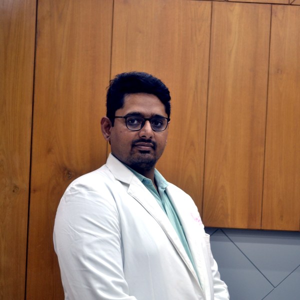
Dr Patel leads deep-endometriosis mapping (expert ultrasound and MRI correlation) and multidisciplinary excision with an organ- and ovarian-sparing, ESGE/ESHRE-aligned approach — operating decisively when it is indicated, and avoiding it when it is not.
Where deep disease sits — and what it causes
| Site | Typical symptom |
|---|---|
| Uterosacral ligaments | Deep pain with intercourse |
| Rectovaginal septum | Painful bowel movements |
| Bowel | Cyclical bowel pain or bleeding |
| Bladder | Cyclical painful urination |
| Ureter | Risk of silent obstruction |
Frequently asked questions
What is deep infiltrating endometriosis?
Deep infiltrating endometriosis (DIE) is endometriosis that extends more than 5 mm beneath the peritoneal surface, most often involving the uterosacral ligaments, rectovaginal space, bowel, bladder or ureters. It is the most complex form of the disease, and it is best managed with precise imaging-based mapping and an individualised, fertility-aware plan.
How is deep infiltrating endometriosis diagnosed — do I need an operation first?
No. Following the ESHRE 2022 guideline, expert transvaginal ultrasound — with MRI where needed — can identify and map most deep disease, so treatment can be planned without a diagnostic operation. One caveat is honest: superficial disease can be invisible on imaging, so a normal scan with persistent symptoms still deserves follow-up rather than dismissal.
Can deep infiltrating endometriosis be treated without surgery?
Often, yes. Hormonal therapy is first-line for pain in many patients and can control symptoms long-term. The choice depends on your pain burden, fertility goals, disease map and how you respond — DIE managed medically still needs structured follow-up, because the aim is long-term disease control, not a one-time fix.
When is surgery genuinely indicated for DIE?
Surgery is indicated when symptoms or organ function do not respond to medical management, when the bowel, bladder or ureters are threatened, or when it serves a specific fertility plan. When indicated, it should be planned around the imaging map by an experienced multidisciplinary team — precise, purposeful and tissue-sparing, with the pelvic anatomy and ovarian reserve protected.
Does deep infiltrating endometriosis affect fertility — and should IVF come before surgery?
DIE can affect fertility, but neither surgery nor IVF is universally first. Sequencing depends on age, ovarian reserve, pain, disease distribution and how much time you have. Repeated ovarian surgery costs reserve, so the question is never only whether an operation helps now, but what it forecloses later — this is exactly what an individualised consultation works through.
Accurate mapping of bowel, bladder and rectovaginal involvement is what makes one definitive, organ-sparing operation possible.
Related reading
Deep infiltrating endometriosis (DIE) requires a thorough preoperative workup to map disease extent and a multidisciplinary surgical team. Conservative management has limited efficacy when there is anatomical distortion or organ involvement.
CONTINUE READING
Explore the Endometriosis Programme
Deep Infiltrating Endometriosis is one part of the broader endometriosis programme led by Dr. Priyadatt Patel. The main endometriosis pillar covers diagnosis, individualised treatment, fertility implications, and long-term management. Related: Deep Infiltrating · 7-10 Year Diagnostic Delay · Multidisciplinary Surgery.
Free Patient Guide
The Endometriosis Decision Guide
A short clinical primer on diagnostic delay, the four decisions you may face, and what to bring to your specialist consultation. Aligned with ESHRE 2022, ASRM, FIGO guidance.
Reviewed by Dr. Priyadatt Patel — read in 20–25 minutes
Free — delivered to your inbox
When surgery is indicated. Deep or recurrent endometriosis sometimes needs operative treatment. Where excision is clearly indicated, see our approach to advanced fertility-preserving laparoscopic excision in Ahmedabad — chosen only on clear indication, prioritising ovarian-reserve preservation.
Dr. Priyadatt Patel
Senior Gynecologist · Advanced Laparoscopic Surgeon · IVF and Endometriosis Programme Lead
MS OBGyn · Pregnancy Care · Advanced Gynaecological Ultrasound · Fertility Preservation
ESHRE / ESGE / AAGL / ASRM guideline-aligned practice. 3D Karl Storz precision technique. Fertility-preservation-first philosophy. Evidence-based decisions, honest counselling, long-term outcomes orientation.
323, Satyamev Eminence, Beside Saptak Bungalows & AUDA Water Tank, Science City Road, Sola, Ahmedabad 380060
Open 24 hours (emergencies & labour) · OPD Mon–Sat 11:00–20:00 · +91 97234 31544
Naranpura, Ahmedabad
Mon–Sat 08:30–10:30 · +91 70460 02566