
Endometriosis · Institutional position
Why endometriosis diagnosis takes 7 to 10 years — and how to shorten it
The average global delay between first symptoms and a confirmed diagnosis of endometriosis is between 7 and 10 years. That figure is not a curiosity. It is a measurable cost — in pain that goes untreated, in fertility windows that close, in mental health, in disease that progresses, and in surgery that becomes more complex than it needed to be. This page sets out the reasons for the delay, the institutional position on it, and the practical steps that can shorten it.
The scale of the delay
Studies from Europe, North America, Australia, and South Asia consistently report a diagnostic delay in endometriosis of 7 to 10 years, sometimes longer. The delay is greatest in two groups: adolescents whose period pain is normalised, and women whose symptoms are predominantly extra-pelvic (bowel, bladder, fatigue) and are therefore diagnosed with something else first.
This is not a quirk of any particular health system. It is a pattern across high-income and middle-income countries alike.
Why the delay happens
- Normalisation of period pain. Severe dysmenorrhoea is often treated as “just bad periods” by family, peers, and clinicians. The patient learns to expect pain rather than to investigate it.
- Symptom heterogeneity. Endometriosis presents with a wide range of symptoms — pelvic pain, bowel symptoms, bladder symptoms, fatigue, painful intercourse, subfertility. No single symptom is pathognomonic.
- Imaging limitations. Endometriosis does not always show on transvaginal ultrasound or MRI — particularly superficial peritoneal disease. A normal scan does not exclude the diagnosis. Many patients are reassured by a normal scan and lose another year before someone questions it.
- The “no laparoscopy without imaging” trap. Some pathways require imaging confirmation before laparoscopy, which excludes patients with surgically-visible-but-imaging-negative disease.
- Differential diagnoses that mimic. Irritable bowel syndrome, interstitial cystitis, pelvic-floor dysfunction, primary dysmenorrhoea — all can coexist with or be misdiagnosed instead of endometriosis.
- Symptom under-reporting. Patients describe their worst symptoms; clinicians may not ask about cyclical pattern, dyspareunia, bowel-bladder symptoms.
- System factors. Short consultation time, sequential referrals between specialists, and a lack of single-point coordination all extend the timeline.
What the delay costs the patient
- Pain. Years of pain that could have been treated.
- Fertility. Years of declining ovarian reserve, lost windows for cycle planning, more demanding IVF later.
- Disease progression. Superficial disease may progress to deep infiltrating disease; ovarian endometriomas may grow.
- Mental-health burden. Anxiety, depression, isolation, and damage to relationships and careers.
- Surgical complexity. Late-diagnosed disease often requires more extensive surgery than early-diagnosed disease would have.
- Cost. Years of investigation, sequential specialists, and trial treatments add to direct and indirect cost.
What shortens the delay
Several practical steps shorten the diagnostic pathway:
- Take period pain seriously. Severe dysmenorrhoea that limits function is not normal. It is a clinical sign that warrants evaluation.
- Document the symptom pattern. A simple cycle diary recording pain location, intensity, bowel-bladder symptoms, and dyspareunia, kept for 2 to 3 cycles, is a more powerful diagnostic tool than many clinicians realise.
- Use ISUOG IDEA-protocol ultrasound. A structured systematic scan is far more sensitive for deep infiltrating disease than a generic pelvic ultrasound. Insisting on the IDEA protocol when arranging imaging is reasonable.
- Trial of medical therapy without delay. Where suspicion is reasonable and imaging is reassuring, an empirical trial of hormonal therapy is appropriate. A clear response supports the diagnosis; failure prompts further evaluation.
- Refer earlier rather than later. A patient whose symptoms have not improved with 6 to 12 months of first-line medical therapy benefits from a specialist opinion. Waiting longer rarely helps.
- Ask the right questions. Clinicians who explicitly ask about cyclical pattern, deep dyspareunia, dyschezia, and cyclical urinary symptoms catch more cases.
- Trust the patient’s account. The patient knows what is normal for her body. Persistent reports of severe symptoms warrant investigation even when investigations are equivocal.
The institutional position
This centre approaches endometriosis as a disease of delayed diagnosis. The first consultation is structured to elicit the full symptom history rather than to dismiss it. Imaging is performed to ISUOG IDEA standards. Medical therapy trials are paced with explicit review points rather than left open-ended. Laparoscopy is reserved for clear indications, not used as a primary diagnostic for surface disease that is better managed medically. Where surgery is the right answer, it is performed by an experienced operator with multidisciplinary backup. And second opinions — ours or anywhere else — are welcomed, not discouraged.
What patients can do today
- Keep a simple cycle diary for 2 to 3 cycles before the consultation
- Note the worst sites of pain, the worst times in the cycle, and the impact on daily life
- List the medications already tried — with doses, durations, and response
- Bring prior ultrasound and MRI reports with images on a USB or disc
- Bring a list of questions to the consultation
- Bring a support person if useful
What clinicians can do
- Ask about cyclical pattern in any patient with pelvic, bowel, or urinary symptoms
- Use the ISUOG IDEA protocol for pelvic ultrasound
- Refer at the 6 to 12-month mark when first-line medical therapy has not relieved symptoms
- Avoid the trap of “normal scan, no further action”
- Document fertility goals at the first visit
- Coordinate care rather than refer sequentially through multiple specialists
Guidelines we follow on this topic
- ESHRE Endometriosis Guideline 2022
- World Endometriosis Society consensus on diagnosis
- RCOG/BSGE guidance
- NICE NG73 Endometriosis
Related reading
The average diagnostic delay for endometriosis remains 7-10 years from symptom onset to definitive diagnosis. Reducing this delay requires high clinical suspicion in primary care and direct access pathways to specialist evaluation.
CONTINUE READING
Explore the Endometriosis Programme
Endometriosis Diagnostic Delay is one part of the broader endometriosis programme led by Dr. Priyadatt Patel. The main endometriosis pillar covers diagnosis, individualised treatment, fertility implications, and long-term management. Related: Deep Infiltrating · 7-10 Year Diagnostic Delay · Multidisciplinary Surgery.
Free Patient Guide
The Endometriosis Decision Guide
A short clinical primer on diagnostic delay, the four decisions you may face, and what to bring to your specialist consultation. Aligned with ESHRE 2022, ASRM, FIGO guidance.
Reviewed by Dr. Priyadatt Patel — read in 20–25 minutes
Free — delivered to your inbox
Why diagnosis takes 7–10 years
| Cause | Effect |
|---|---|
| Normalised period pain | Late presentation |
| Non-specific symptoms | Misattribution (IBS, etc.) |
| Negative scans | False reassurance |
| Fragmented care | The pattern is missed |
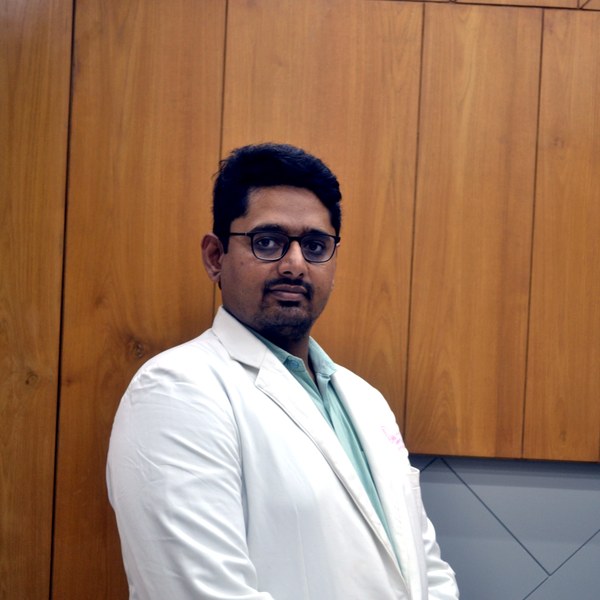
Dr Patel leads endometriosis diagnosis and surgery at Balaji Horizon with an evidence-based, ovarian-sparing philosophy aligned to ESHRE and ESGE — integrating pain, fertility and long-term disease control into a single plan, rather than treating the disease in isolation.
Imaging-led diagnosis, medical-first management, and precise surgery only when it is the right step — planned around your pain and fertility goals.
Dr. Priyadatt Patel
Senior Gynecologist · Advanced Laparoscopic Surgeon · IVF and Endometriosis Programme Lead
MS OBGyn · Pregnancy Care · Advanced Gynaecological Ultrasound · Fertility Preservation
ESHRE / ESGE / AAGL / ASRM guideline-aligned practice. 3D Karl Storz precision technique. Fertility-preservation-first philosophy. Evidence-based decisions, honest counselling, long-term outcomes orientation.
323, Satyamev Eminence, Beside Saptak Bungalows & AUDA Water Tank, Science City Road, Sola, Ahmedabad 380060
Open 24 hours (emergencies & labour) · OPD Mon–Sat 11:00–20:00 · +91 97234 31544
Naranpura, Ahmedabad
Mon–Sat 08:30–10:30 · +91 70460 02566