
How long does the anomaly scan take?
The scan itself usually takes about 30 to 45 minutes. It can take longer when the baby’s position hides some views, and occasionally a short repeat look is arranged on another day to complete them. A longer scan, or a repeat look, does not by itself mean that something is wrong.
Anomaly Scan (TIFFA) in Pregnancy — Your Detailed 18–22 Week Fetal Survey
The anomaly scan, or TIFFA (Targeted Imaging for Fetal Anomalies), is the most detailed ultrasound of your pregnancy — a careful, structured check of your baby’s growth and development. At Balaji Horizon it is performed to ISUOG-aligned standards on advanced GE Voluson™ S10, P6 and Swift systems.
In short
The anomaly scan — also called TIFFA — is a detailed ultrasound performed between 18 and 22 weeks. It examines the baby’s brain, heart, spine, face, limbs and abdominal organs, along with the placenta, cord and amniotic fluid. It screens for structural differences and cannot detect every condition. Fetal sex is neither determined nor disclosed, as per the PC-PNDT Act.
What is the anomaly scan — and why does it matter?
The anomaly scan is a detailed, systematic ultrasound performed in the middle of pregnancy to examine your baby’s anatomy organ by organ. Its purpose is twofold: to offer reassurance that your baby is developing as expected, and to identify the small number of babies who may benefit from closer monitoring, specialist input or a clear plan for delivery.
One scan, many names. “TIFFA scan”, “anomaly scan”, “Level II scan” and the “20-week scan” all describe the same examination — TIFFA simply stands for Targeted Imaging for Fetal Anomalies. Different doctors and regions use different names for it.
When is the TIFFA scan done, and why that window?
The anomaly scan is best performed between 18 and 22 weeks of pregnancy, and we typically schedule it at 19–21 weeks. This window is chosen carefully: by now the baby’s organs are fully formed and large enough to be examined in detail, yet there is still adequate time to arrange further assessment or planning if it is ever needed.
If the baby is lying in an awkward position — which is common — we may ask you to walk for a few minutes, have something to eat, or return for a short follow-up. This is routine and never a cause for concern; a complete, unhurried survey always takes priority over speed.
The complete systematic survey: what we examine
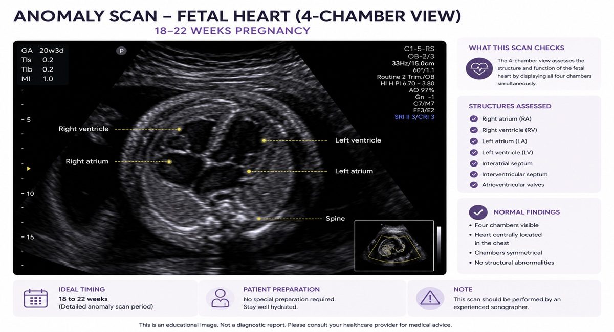
An anomaly scan is not a single picture — it is a structured, head-to-toe protocol covering every major organ system. Below is what a thorough examination reviews, and the kinds of conditions each part of the survey can help to identify. A system-by-system account of what is examined, and what each part of the survey is looking for, is set out in every organ system the anomaly scan checks. Where a cardiac question arises, fetal echocardiography explains when a dedicated heart study is indicated.
| System examined | What is assessed | Examples it can help detect |
|---|---|---|
| Head & brain | Skull shape, ventricles, cavum septi pellucidi, cerebellum, cisterna magna | Anencephaly, ventriculomegaly, Dandy-Walker malformation, cranial signs of spina bifida |
| Face | Orbits, upper lip, facial profile, nasal bone | Cleft lip (and many cleft-palate cases) |
| Spine | Full length in long and transverse planes, overlying skin | Open spina bifida, hemivertebrae, scoliosis |
| Heart | Four-chamber view, left & right outflow tracts, three-vessel & trachea view, rate and rhythm | Many major congenital heart defects |
| Chest & lungs | Lung appearance, diaphragm, heart position | Diaphragmatic hernia, lung lesions (e.g. CPAM), effusions |
| Abdomen | Stomach, bowel, abdominal wall, cord insertion | Gastroschisis, exomphalos, bowel obstruction |
| Kidneys & bladder | Both kidneys, renal pelvis, bladder filling | Renal agenesis, significant hydronephrosis, obstruction |
| Limbs | Both arms and legs, long bones, hands and feet | Skeletal dysplasia, limb-reduction defects, talipes (club foot) |
| Placenta, cord & fluid | Placental position, number of cord vessels, amniotic fluid volume | Low-lying placenta, single umbilical artery, abnormal fluid |
| Growth (biometry) | Head, abdomen and femur measurements | Confirms the baby is an appropriate size for dates |
- A defined sequence is followed every time, so no view is missed.
- Key images and measurements are stored and documented in a structured report.
- 3D/4D imaging is used selectively where it adds clinical value — not as a substitute for the standard 2D survey.
What the anomaly scan can — and cannot — detect
Honesty matters here. The anomaly scan is a powerful screening test, but it is a screening test — not a guarantee. Detection depends on the specific condition, the baby’s position, maternal factors and the timing of the scan. The figures below are approximate sensitivities reported in the literature and serve as a realistic guide, not a promise.
| Condition | Approximate detection | Notes |
|---|---|---|
| Anencephaly / acrania | ~97–100% | Often already visible on the first-trimester scan |
| Open spina bifida | ~90% | Aided by characteristic cranial signs |
| Abdominal wall defects | ~90% | Gastroschisis, exomphalos |
| Major kidney anomalies | ~85–90% | e.g. bilateral renal agenesis |
| Major congenital heart disease | ~50–60% | Higher with outflow-tract and 3-vessel views in expert hands |
| Diaphragmatic hernia | ~50–60% | Can be subtle in mid-pregnancy |
| Limb-reduction defects | ~60–75% | Depends on type and severity |
Soft markers — and why most are nothing to fear

A “soft marker” is a minor, often temporary finding that is common in perfectly healthy babies. In isolation, in a low-risk pregnancy, most soft markers carry little or no significance. Their meaning comes from context — your screening results, whether more than one marker is present, and the overall picture.
| Soft marker | What it is | Usual significance (isolated, low-risk) |
|---|---|---|
| Echogenic intracardiac focus | A bright spot in the heart | Very common; usually no significance |
| Choroid plexus cyst | A small cyst in the brain’s fluid tissue | Almost always resolves; benign in isolation |
| Mild renal pelvis dilatation | Slightly prominent kidney drainage | Often a follow-up scan in the third trimester |
| Echogenic bowel | Brighter-than-usual bowel | Reviewed in context of other findings |
| Short femur / humerus | Slightly short long bones | Interpreted with growth and screening |
| Single umbilical artery | Two cord vessels instead of three | May prompt growth monitoring |
Beyond anatomy: what else the scan assesses
A thorough mid-pregnancy scan looks after the pregnancy as a whole, not only the baby’s structure:
- Placental position — checking it is clear of the cervix, with a follow-up later if it is low-lying.
- Amniotic fluid volume — confirming a healthy environment for your baby.
- Cervical assessment — where indicated, to help identify women who may benefit from preterm-birth prevention.
- Growth and wellbeing — biometry to confirm appropriate size, with Doppler studies added when clinically indicated.
If a finding needs a closer look
Most anomaly scans are reassuring. When something does need further evaluation, it rarely means bad news — it means we look more carefully and plan thoughtfully. Depending on the finding, the next step may be:
- A targeted repeat scan after a short interval, often resolving the question entirely.
- Fetal echocardiography — a detailed study of the baby’s heart.
- A neurosonogram — dedicated imaging of the brain.
- Fetal MRI in selected cases, as a complement to ultrasound.
- Counselling about invasive testing (such as amniocentesis) where a genetic question arises — always your informed choice.
- Multidisciplinary fetal-medicine input and, where helpful, genetic counselling.
How the anomaly scan fits with your other scans
The pregnancy scans are complementary — each answers a different question at a different stage.
| Scan | Timing | Main purpose |
|---|---|---|
| NT scan + screening | 11–13+6 weeks | Chromosomal risk screening (Down syndrome and others) |
| Anomaly scan (TIFFA) | 18–22 weeks | Detailed structural survey of the baby’s anatomy |
| Growth scan | Third trimester | Growth, fluid and wellbeing before delivery |
The technology behind your anomaly scan
In fetal medicine, subtle findings are only as clear as the system resolving them. Your anomaly scan is performed on GE Voluson™ platforms across our two locations, with advanced 3D/4D and AI-assisted imaging used where it adds genuine clinical value.
Voluson S10 Expert XD
Our flagship — Voluson Core Architecture with HDlive™ and Radiantflow™ rendering and XD Clear™ single-crystal transducers (C2-9-RS & RAB6-RS), plus advanced fetal-cardiac tools (STIC) and AI Sono-automation, for exceptional anatomical and blood-flow detail.
Voluson SWIFT Plus
AI-assisted imaging with SonoLyst image recognition and a fast, ergonomic workflow — consistent, high-quality views with a smoother examination.
Voluson P6
A reliable, full-featured obstetric system with 2D/3D/4D imaging — complete early-pregnancy and anomaly assessment at our second location.
3D and 4D imaging. Beyond the standard 2D survey, 3D builds a sculpted view of structures such as the face and spine, while 4D shows your baby moving in real time. On our S10 Expert, HDlive™ and Radiantflow™ produce strikingly realistic surface and blood-flow imaging. We use them selectively — to clarify a finding or share a reassuring moment — never as a substitute for the systematic diagnostic survey. See our 3D/4D scan for keepsake imaging.
Images from our Voluson S10 Expert
A few examples of the imaging this system produces in everyday fetal-medicine practice:
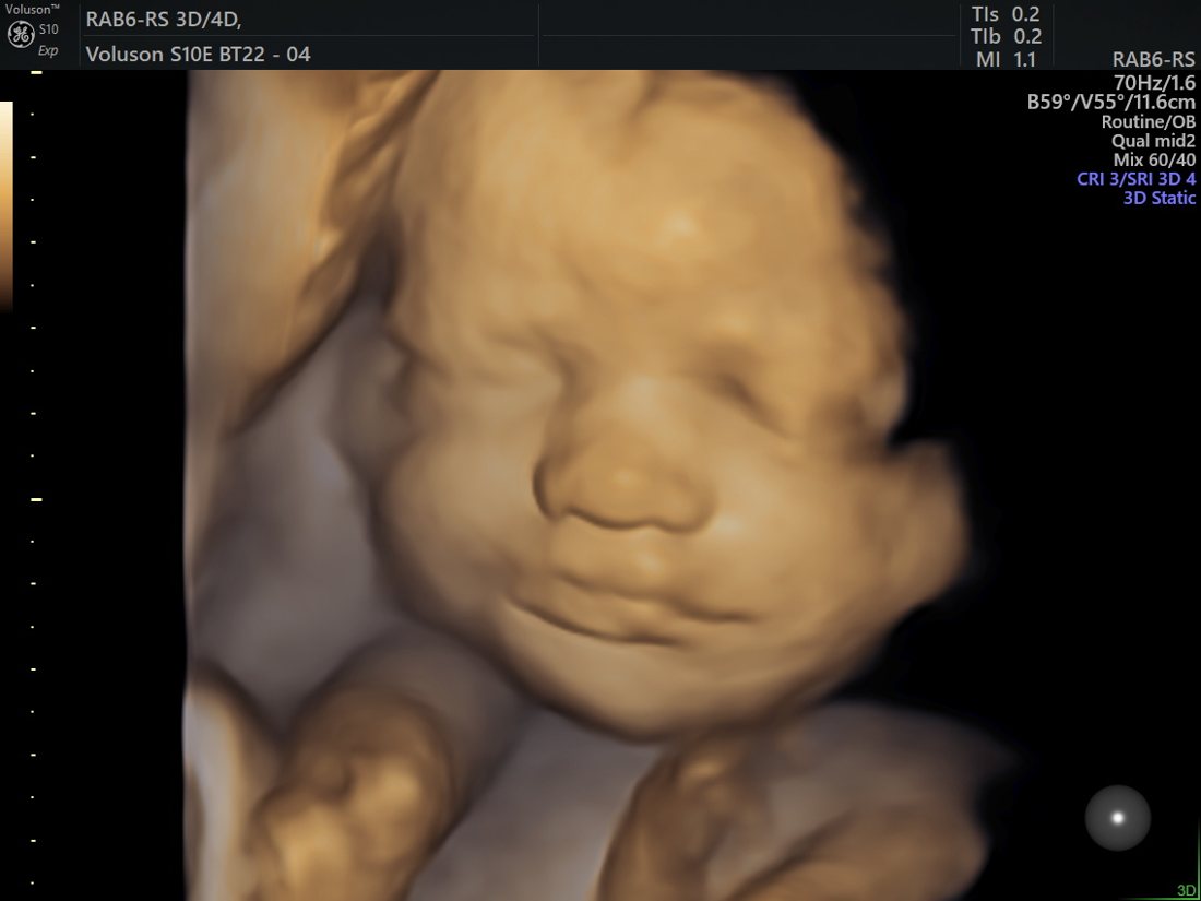
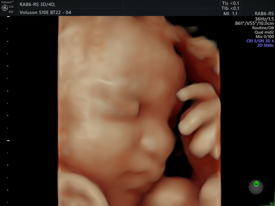
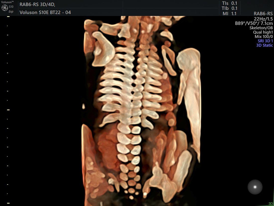
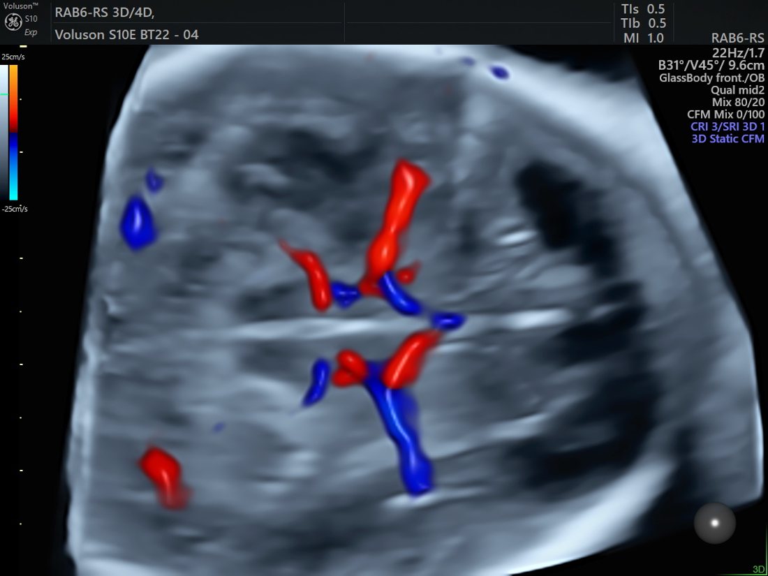
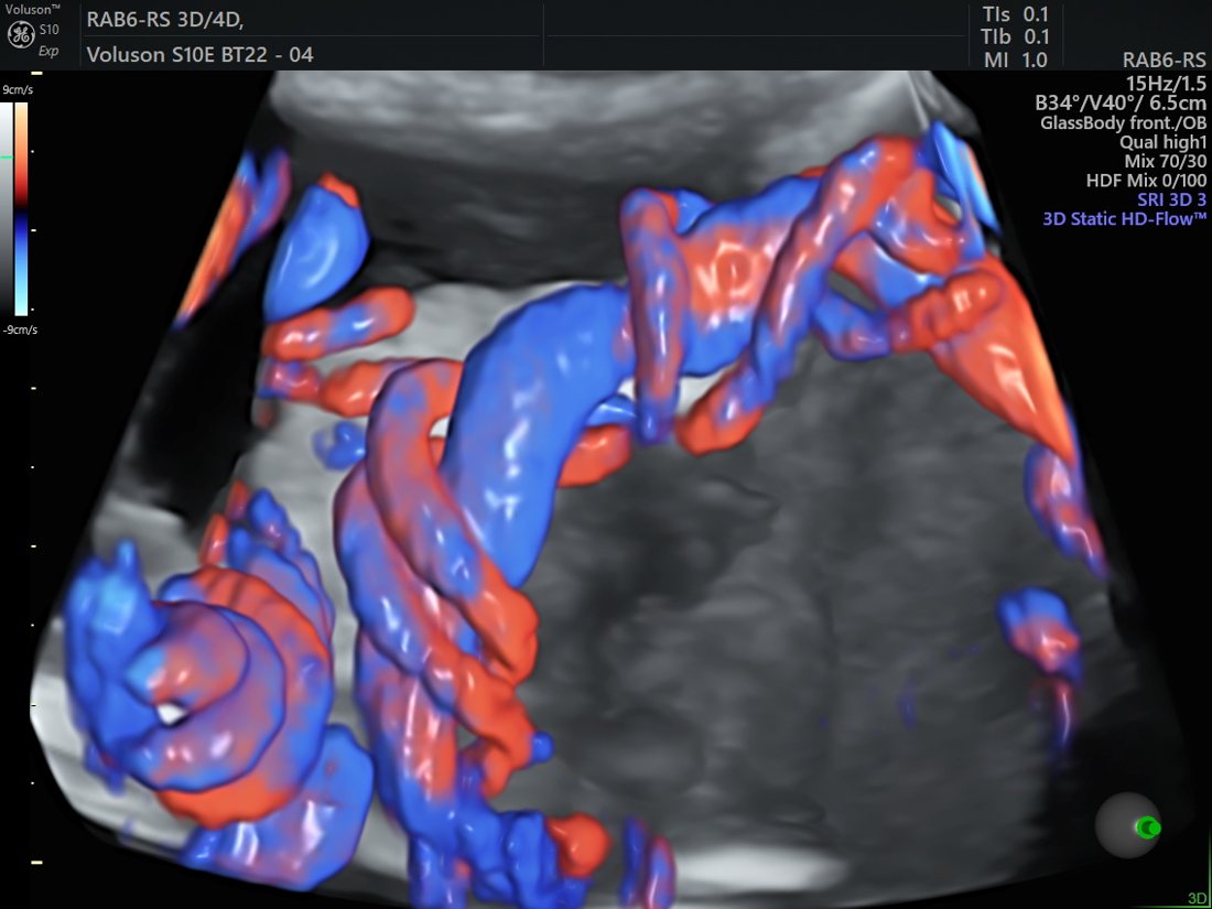
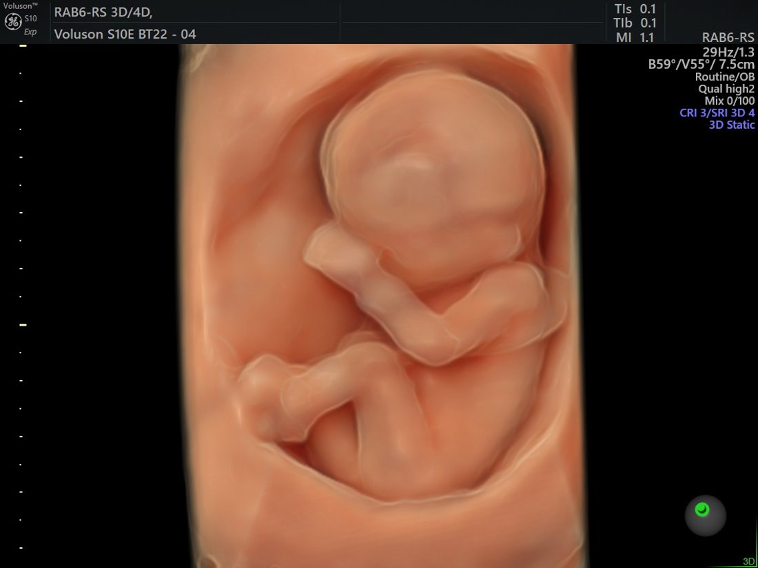
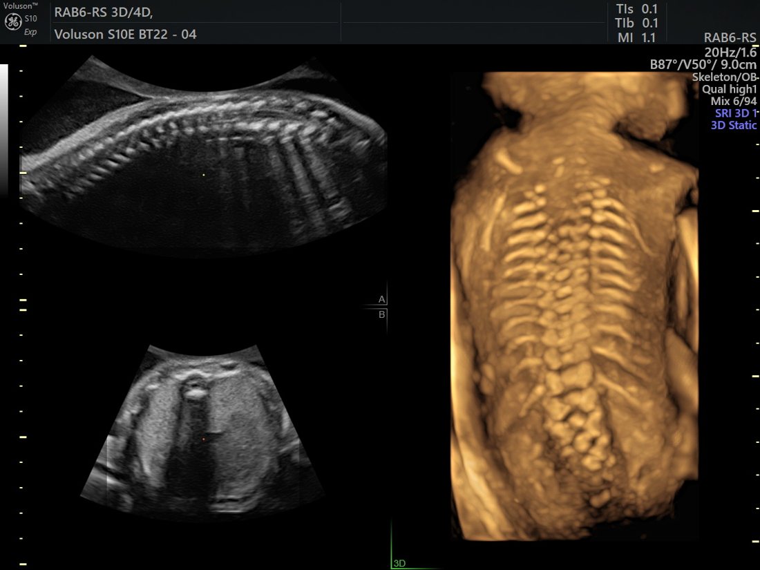
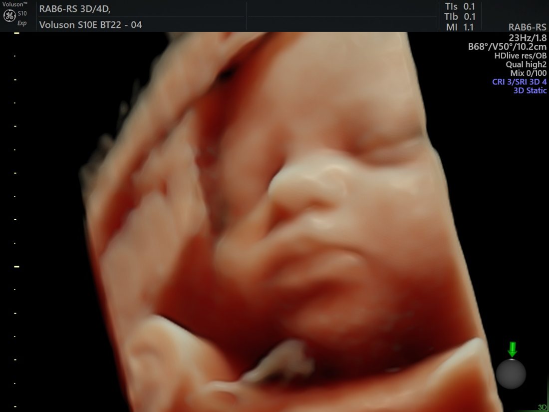
Actual images acquired on our GE Voluson™ S10 Expert. “GE”, “Voluson”, “HDlive” and “Radiantflow” are trademarks of GE HealthCare; shown with permission.
Your fetal-medicine team
Your anomaly scan is performed and interpreted by a clinician dedicated to fetal medicine, with a direct pathway to senior gynaecologist Dr. Priyadatt Patel for any pregnancy that needs obstetric care.

Dr. Mayank Chaudhary
An ISUOG-trained fetal-medicine consultant who performs the detailed anatomy survey to international standards — anomaly scanning, fetal cardiac assessment and high-risk pregnancy imaging.
Your anomaly scan at Balaji Horizon, Ahmedabad
Our fetal-medicine service is built around accuracy, clarity and calm:
- Advanced imaging. Scans are performed on GE Voluson™ S10, P6 and Swift systems, with AI-assisted 3D/4D capability used where it adds clinical value.
- ISUOG-aligned protocol. A structured, internationally benchmarked survey with documented images and measurements.
- Specialist interpretation. Reviewed by clinicians with dedicated fetal-medicine and advanced-ultrasound expertise.
- Clear, structured reporting. You receive a thorough report and a plain-language explanation of what it means.
- Continuity of care. If a finding needs more, the same team guides you through every next step.
The anomaly scan in your pregnancy scan schedule
The anomaly scan — also called the TIFFA or 20-week scan — is a detailed mid-pregnancy ultrasound, performed between 18 and 22 weeks, that systematically examines your baby’s anatomy from head to toe. It is one of the two routine scans of an uncomplicated pregnancy, alongside the first-trimester dating and NT scan.
To see how the anomaly scan fits with every other scan across all three trimesters — which scan, and when — read the full pregnancy ultrasound scan schedule.
Your TIFFA report — what it contains and how to read it
Most women leave the scan room with a printed report and very little idea of how to read it. The structure is the same everywhere, and knowing it makes the report far less intimidating.
- Dating and basics. The gestational age used for comparison, the number of babies, cardiac activity, and how your baby was lying.
- Biometry. Head, abdominal and thigh-bone measurements, converted into an estimated fetal weight and a percentile. A percentile is a position in a range, not a mark out of a hundred.
- The anatomical survey. Each organ system listed in turn — brain, face and lips, spine, heart, abdominal wall, stomach, kidneys, bladder, limbs. Each is reported as appearing normal, or the finding is described in plain terms.
- The environment. Placental position and appearance, amniotic fluid volume, umbilical cord vessels, and cervical length where it is indicated.
- Limitations. An honest note of what could not be seen well — fetal position and maternal body habitus both affect what is visible.
- Recommendation. Whether anything needs a repeat scan, a fetal echocardiogram, or a specialist opinion.
Detection is not uniform across organ systems: some conditions are reliably visible at this gestation, others are not visible at any gestation, and a few declare themselves only later in pregnancy. That is why a normal anomaly scan is reported as reassuring rather than as a clean bill of health — an honest report says what was seen, on that day, under those conditions.
Your report will not state fetal sex. Fetal sex is neither determined nor disclosed, as per the PC-PNDT Act.
Frequently asked questions
In which week should the anomaly / TIFFA scan be done?
How long does the scan take?
Is the TIFFA scan the same as the anomaly scan?
Do I need to fast or prepare?
Is the scan safe for my baby?
What if the baby is in a difficult position?
Does a normal anomaly scan guarantee a healthy baby?
Can the anomaly scan detect Down syndrome?
Can the scan tell the baby’s sex?
What does the TIFFA scan report contain?
What will I see on the screen during the TIFFA scan?
How do I book the anomaly (TIFFA) scan?
Book your anomaly (TIFFA) scan
A detailed, unhurried 18–22 week survey on advanced GE Voluson imaging, with clear answers from a specialist fetal-medicine team in Ahmedabad.
If the scan raises questions, the next step is usually structured counselling rather than alarm — see genetic counselling and prenatal testing options.
If you are deciding between scans earlier in pregnancy, this comparison may help: NT scan vs anomaly (TIFFA) scan — what each one checks, and when.
Anomaly scan — also known as
Also known as: TIFFA scan · Targeted Imaging For Fetal Anomalies · Level II scan · 20-week scan · anatomy scan. These all describe this same detailed 18–22 week fetal-anatomy survey. See also the NT scan and NT scan vs anomaly (TIFFA) scan.
Your fetal-medicine specialist
Fetal-medicine imaging at Balaji Horizon is led by Dr. Mayank Chaudhary, an ISUOG-trained fetal medicine specialist & sonologist — performing detailed anomaly (TIFFA) scanning, fetal echocardiography and high-risk pregnancy surveillance with the obstetric team. Meet the team →
Dr. Priyadatt Patel
Senior Gynecologist · Advanced Laparoscopic Surgeon · IVF and Endometriosis Programme Lead
MS OBGyn · Pregnancy Care · Advanced Gynaecological Ultrasound · Fertility Preservation
ESHRE / ESGE / AAGL / ASRM guideline-aligned practice. 3D Karl Storz precision technique. Fertility-preservation-first philosophy. Evidence-based decisions, honest counselling, long-term outcomes orientation.
323, Satyamev Eminence, Beside Saptak Bungalows & AUDA Water Tank, Science City Road, Sola, Ahmedabad 380060
Open 24 hours (emergencies & labour) · OPD Mon–Sat 11:00–20:00 · +91 97234 31544
Naranpura, Ahmedabad
Mon–Sat 08:30–10:30 · +91 70460 02566