
Growth Monitoring • 28–40 Weeks
Fetal Growth Scan — Biometry and Estimated Fetal Weight
In short
A growth scan estimates the baby’s weight from head, abdomen and femur measurements and plots it on a chart. A single measurement matters less than the trend across scans. Estimated weight carries an inherent margin of error, which is why growth is judged over time rather than from one number. Fetal sex is neither determined nor disclosed, as per the PC-PNDT Act.
Accurate fetal size and growth assessment using standardised biometric measurements — monitoring growth trajectory across the third trimester. Science City & Naranpura, Ahmedabad.
What Does a Fetal Growth Scan Assess?
A fetal growth scan measures the baby’s dimensions to assess growth trajectory and estimate fetal weight. Unlike the anomaly scan, which focuses on structural detail, growth scans track whether the baby is growing at the expected rate for its gestational age — and whether that rate is sustained between appointments.
Biometric measurements taken:
Growth Scan Schedule — When and How Often?
| Timing | Purpose | Who Needs It |
|---|---|---|
| 28 weeks | Baseline third trimester assessment — establishes growth trajectory | All pregnancies (especially high-risk) |
| 32 weeks | Mid-third trimester growth velocity — check for late-onset growth restriction | All high-risk; optional in low-risk |
| 36 weeks | Pre-delivery assessment: EFW, presentation, liquor, placenta location | All pregnancies |
| Every 2–3 weeks | Monitoring confirmed or suspected growth restriction | Twins, diabetes, hypertension, prior SGA, FGR |
Understanding Fetal Growth Restriction (FGR)
Fetal Growth Restriction (FGR) — previously termed IUGR — is a condition where the fetus fails to reach its genetic growth potential, most commonly due to placental insufficiency. Early identification and monitoring prevents adverse outcomes.
| Type | Onset | Characteristics | Management |
|---|---|---|---|
| Early-onset FGR | <32 weeks | Often associated with severe placental disease; significant Doppler abnormalities; worse prognosis | Intensive monitoring; timing of delivery critical |
| Late-onset FGR | >32 weeks | More common; subtler Doppler changes; may be missed without serial scanning | Serial growth + Doppler scans; planned delivery |
Key indicators on growth scan:
- ✓AC or EFW below the 10th centile for gestational age
- ✓AC crossing centile lines downward between serial scans
- ✓Reduced or absent amniotic fluid (oligohydramnios)
- ✓Abnormal umbilical artery Doppler waveforms (next step after growth scan)
Growth Scanning at Balaji Horizon
- Precise biometric measurements: GE Voluson systems at both Science City and Naranpura provide the image quality required for accurate HC, AC, FL, and BPD measurements.
- INTERGROWTH-21st and Hadlock growth charts: Measurements are compared against internationally validated growth standards for accurate centile assessment.
- ISUOG-trained specialist: Dr. Mayank Chaudhary performs and interprets all growth assessments to international standards.
- Integrated Doppler assessment: When growth restriction is suspected, umbilical artery and MCA Doppler are performed in the same appointment — avoiding separate visits.
- High-risk obstetric integration: All significant findings feed directly into Dr. Priyadatt Patel’s high-risk obstetric management pathway for planning.
Images from our Voluson S10 Expert
Examples of the biometry and growth imaging this system produces in everyday practice at Balaji Horizon:
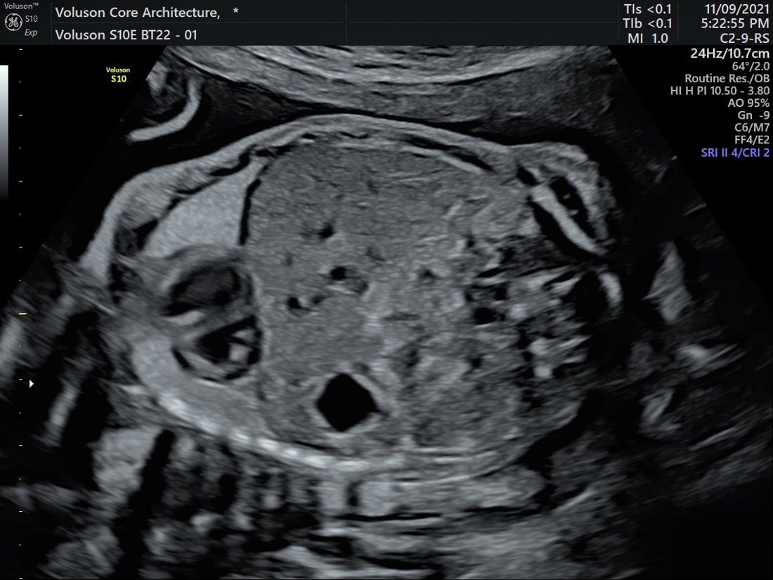
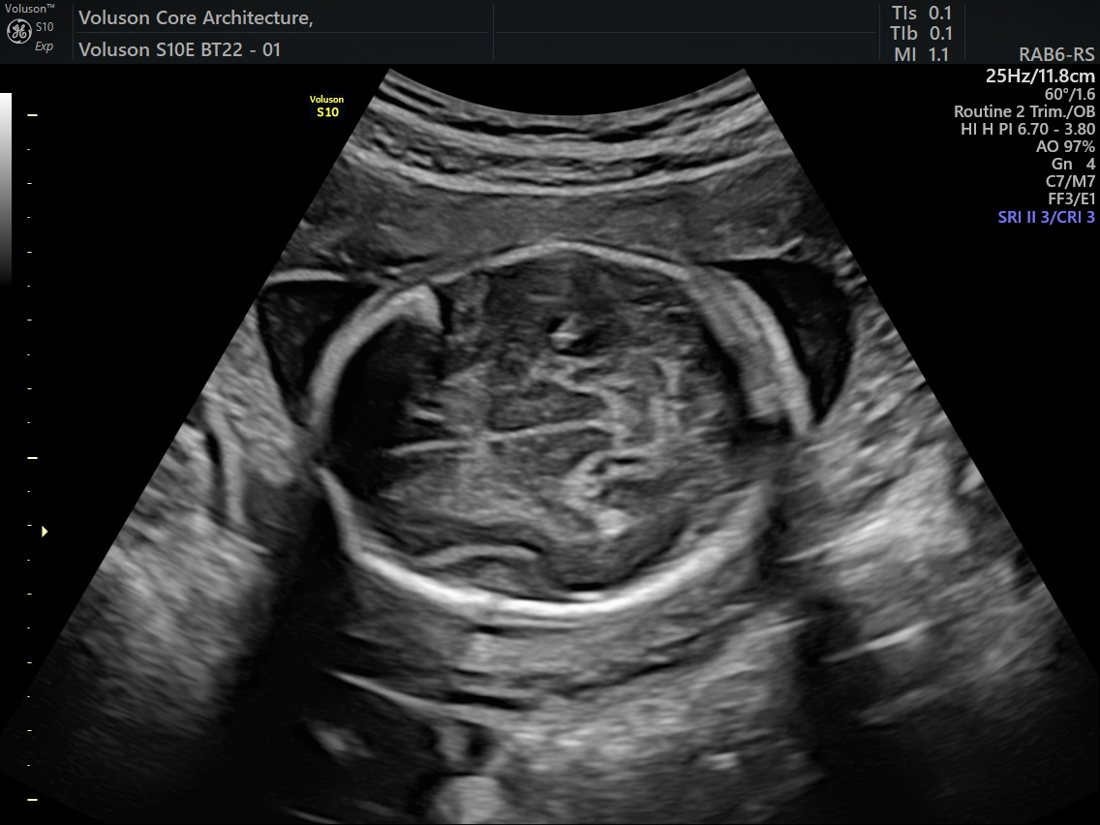
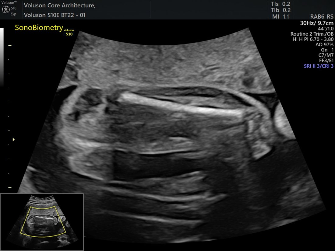
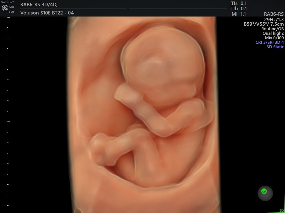
Actual images acquired on our GE Voluson™ S10 Expert. “GE”, “Voluson”, “HDlive” and “Radiantflow” are trademarks of GE HealthCare; shown with permission.
Your fetal-medicine specialist
Your growth scan is performed and interpreted by a clinician dedicated to fetal medicine, with a direct pathway to senior gynaecologist Dr. Priyadatt Patel for any pregnancy that needs obstetric care.

Dr. Mayank Chaudhary
An ISUOG-trained fetal-medicine consultant who performs detailed fetal assessments to international standards.
Standards & further reading. Our protocol aligns with the ISUOG practice guidelines. For independent patient information see the NHS guide to pregnancy ultrasound.
Related fetal scans
How to read a growth-scan result
A growth scan estimates your baby’s weight from head, abdomen and femur measurements and plots it on a percentile chart. A single number is a snapshot — what matters most is the trend across scans and the whole picture.
- Percentiles describe size, not health: a baby on the 10th percentile can be perfectly healthy and simply small. The pattern over time is the key signal.
- The trend matters more than one reading: a falling trend across serial scans is what prompts closer attention — far more than a single low value.
- Read together, not in isolation: the scan also checks amniotic fluid and, when needed, Doppler blood flow; these are interpreted alongside the size.
- Small is not the same as growth restriction: most small (SGA) babies are constitutionally small and well; the aim is to identify the few with true growth restriction (FGR) who need monitoring.
Growth scans are indicated, not routine for everyone — used when there is a reason, such as risk factors, a small or large bump, a previous growth issue, or a medical condition. The goal is to support the pregnancies that need closer watching without over-investigating healthy ones.
See where this fits in your pregnancy: the full pregnancy ultrasound scan schedule, and — where blood flow needs checking — the Doppler scan.
References: ISUOG Practice Guidelines — fetal biometry and growth; NICE NG201, Antenatal care. Educational information, not a substitute for individual medical advice.
Frequently Asked Questions
- Is the estimated fetal weight accurate?EFW has an inherent margin of approximately ±15–20%. A single EFW is less clinically meaningful than a trend across serial scans. Growth velocity — the rate of change in AC and EFW between scans — is more important than a single measurement in isolation.
- My baby is measuring small. Should I be worried?Not necessarily. “Small” can mean constitutionally small (a normal variant for that baby’s genetic make-up) or growth restricted (placental insufficiency). Doppler assessment and growth velocity across serial scans clarify this distinction. Most babies measuring small are entirely healthy.
- How often do I need growth scans?In a low-risk pregnancy: typically at 28, 32, and 36 weeks. In high-risk pregnancies (twins, maternal diabetes, hypertension, prior SGA baby): every 2–3 weeks from 26 weeks, or as directed by your clinician based on findings.
- Can a growth scan detect a large baby (macrosomia)?Yes. EFW above the 90th centile (large for gestational age, LGA) — often associated with maternal diabetes — is identified on growth scan. Management depends on EFW trend, glycaemic control, and clinical context. Macrosomia has implications for mode of delivery planning.
- Does fetal position affect measurement accuracy?Suboptimal fetal positioning can make abdominal circumference (AC) measurement more difficult to obtain accurately. The sonographer will attempt multiple views and optimise measurements. If position is unfavourable, a short wait or repeat appointment may occasionally be required.
Book Your Fetal Growth Scan
Serial growth assessment is essential for high-risk pregnancies. Book your third trimester growth scan at Balaji Horizon to monitor your baby’s growth trajectory with precision.
Related scans:
Frequently asked questions
What is the difference between a growth scan and an anomaly scan?
Is the estimated fetal weight accurate?
My baby is measuring small. Should I be worried?
How often do I need growth scans?
Can a growth scan detect a large baby (macrosomia)?
Does fetal position affect measurement accuracy?
Medically reviewed by Dr. Priyadatt Patel, MBBS, MS (Obstetrics & Gynaecology) — Senior Gynecologist · Advanced Laparoscopic Surgeon · IVF and Endometriosis Programme Lead & Fertility Specialist, Balaji Horizon Women’s Hospital, Ahmedabad. Last reviewed June 2026, aligned with ISUOG fetal-ultrasound standards. This page is for education and does not replace individual medical advice.
Growth scan — also known as
Also known as: growth scan (Ahmedabad) · fetal biometry · third-trimester scan. A growth scan checks your baby's growth and wellbeing later in pregnancy and is different from the mid-pregnancy anomaly scan; when growth is a concern it is paired with a Doppler study.
Dr. Priyadatt Patel
Senior Gynecologist · Advanced Laparoscopic Surgeon · IVF and Endometriosis Programme Lead
MS OBGyn · Pregnancy Care · Advanced Gynaecological Ultrasound · Fertility Preservation
ESHRE / ESGE / AAGL / ASRM guideline-aligned practice. 3D Karl Storz precision technique. Fertility-preservation-first philosophy. Evidence-based decisions, honest counselling, long-term outcomes orientation.
323, Satyamev Eminence, Beside Saptak Bungalows & AUDA Water Tank, Science City Road, Sola, Ahmedabad 380060
Open 24 hours (emergencies & labour) · OPD Mon–Sat 11:00–20:00 · +91 97234 31544
Naranpura, Ahmedabad
Mon–Sat 08:30–10:30 · +91 70460 02566
Growth scan vs anomaly scan — what is the difference?
They answer different questions and are done at different times. The anomaly scan (TIFFA), at about 18–22 weeks, is a detailed one-off survey of the baby’s structure — brain, heart, spine, kidneys, limbs. The growth scan, usually from about 28–32 weeks onward and often repeated, measures size and wellbeing — head, abdomen and femur (biometry) to estimate fetal weight, plus liquor and, where indicated, Doppler blood flow. In short: the anomaly scan asks “is the baby formed normally?”; the growth scan asks “is the baby growing and coping well?” A normal anomaly scan does not remove the need for growth surveillance later, because growth restriction typically develops in the third trimester.
- Anomaly / TIFFA scan — 18–22 weeks · structure · usually once. Read about the anomaly scan.
- Growth scan — from ~28 weeks · size, liquor, Doppler · may be repeated. This page.
- Doppler study — blood flow, added when growth or wellbeing is in question. Read about Doppler.
- Growth surveillance is intensified in a high-risk pregnancy, and a change in movements always needs same-day review — see reduced fetal movements.