
MRI Mapping for Deep Endometriosis — Surgical Planning
MRI complements expert ultrasound for endometriosis diagnosis, particularly for deep infiltrating disease involving bowel, bladder and ureters. This page explains when MRI is used, what it shows, how findings inform surgical planning, and what to expect during the scan.
1. When MRI is indicated
Deep infiltrating endometriosis identified or suspected on ultrasound. Bowel involvement requiring depth and distance measurement for surgical planning. Bladder endometriosis. Suspected ureteric involvement. Pre-operative mapping before complex endometriosis excision. Equivocal ultrasound findings. Frozen pelvis with severe adhesions on dynamic ultrasound.
2. Standard MRI sequences
T2-weighted sequences in three planes (axial, sagittal, coronal) — primary detection of endometriosis as hypointense lesions. T1-weighted with fat saturation — detection of haemorrhagic foci within lesions (highly specific for endometriosis). Diffusion-weighted imaging in selected cases. Standard pelvic MRI takes 30–45 minutes.
3. Special preparations and contrast
Rectal contrast (sonohysterography-like rectal water or gel) improves detection of bowel wall involvement and quantifies lesion depth. Vaginal contrast improves visualisation of posterior fornix and rectovaginal septum. Bowel preparation (laxative or enema) optional but improves image quality. MRI urography for suspected ureteric disease.
4. What MRI shows that ultrasound may not
Exact depth of bowel wall infiltration (mucosal, submucosal, muscularis propria, transmural). Distance from anal verge — critical for deciding shaving vs discoid vs segmental resection. Bilateral ureteric tracking. Diaphragmatic implants when extended chest sequences added. Larger-volume disease mapping for complex multi-compartment cases.
5. Findings on MRI
Endometriosis nodules appear as hypointense (dark) lesions on T2-weighted images, often with internal T1-hyperintense foci (bright spots) representing haemorrhagic content. Endometriomas show characteristic T1 hyperintensity with T2 shading. Adenomyosis shows thickened junctional zone (over 12 mm) and myometrial cysts.
6. Surgical planning value
MRI findings directly inform surgical strategy. Distance of bowel lesion from anal verge: less than 5 cm needs colorectal team for low anterior resection; 5–15 cm allows discoid or shaving; over 15 cm allows simpler approaches. Bilateral ureteric mapping prevents intraoperative surprises. Pre-operative bowel preparation planned where indicated.
7. What to expect during the scan
30–45 minutes lying supine in MRI scanner. No contrast injection in most cases (oral or rectal contrast only). Loud knocking sounds during sequences (earplugs provided). Patient communication via intercom. Mildly claustrophobic for some patients — open MRI alternative available in some centres but with lower image quality.
8. Limitations of MRI
Cost and accessibility higher than ultrasound. May miss small superficial peritoneal lesions. Reader expertise matters — endometriosis MRI interpretation requires specific training. Pacemakers and certain metal implants contraindicate MRI. Pregnancy is not a contraindication but is usually deferred unless urgent.
When MRI adds value
| Indication | What MRI clarifies |
|---|---|
| Deep infiltrating disease | Extent, bowel/bladder depth |
| Surgical planning | A lesion map for the team |
| Large or complex endometriomas | Characterisation |
| Suspected ureteric involvement | Obstruction risk |
Aligned with current international evidence, not habit.
Frequently Asked Questions
Do I need MRI for endometriosis?
How long does MRI take?
Is MRI uncomfortable?
Will I need contrast injection?
Should I have ultrasound or MRI first?
How accurate is MRI for endometriosis?
Can MRI replace ultrasound?
What if I have a pacemaker?
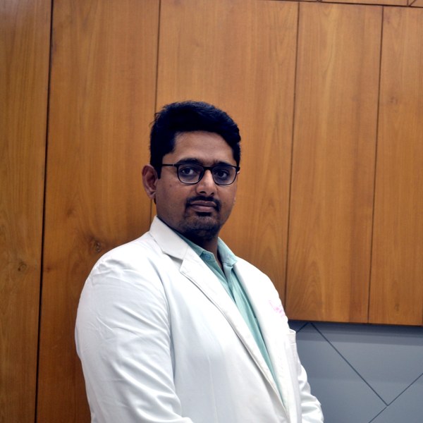
Dr Patel leads endometriosis diagnosis and surgery at Balaji Horizon with an evidence-based, ovarian-sparing philosophy aligned to ESHRE and ESGE — integrating pain, fertility and long-term disease control into a single plan, rather than treating the disease in isolation.
Imaging-led diagnosis, medical-first management, and precise surgery only when it is the right step — planned around your pain and fertility goals.
Dr. Priyadatt Patel
Senior Gynecologist · Advanced Laparoscopic Surgeon · IVF and Endometriosis Programme Lead
MS OBGyn · Pregnancy Care · Advanced Gynaecological Ultrasound · Fertility Preservation
ESHRE / ESGE / AAGL / ASRM guideline-aligned practice. 3D Karl Storz precision technique. Fertility-preservation-first philosophy. Evidence-based decisions, honest counselling, long-term outcomes orientation.
323, Satyamev Eminence, Beside Saptak Bungalows & AUDA Water Tank, Science City Road, Sola, Ahmedabad 380060
Open 24 hours (emergencies & labour) · OPD Mon–Sat 11:00–20:00 · +91 97234 31544
Naranpura, Ahmedabad
Mon–Sat 08:30–10:30 · +91 70460 02566