
Endometriosis · Bowel involvement
Bowel endometriosis — when endometriosis crosses into the bowel
Bowel endometriosis is a recognised form of deep infiltrating disease that involves the wall of the rectum, sigmoid, or, less commonly, the small bowel, caecum, or appendix. It is often missed because the symptoms can mimic irritable bowel syndrome. This page explains how it presents, how it is diagnosed, and how it is managed within a multidisciplinary surgical framework.
What bowel endometriosis is
Bowel endometriosis is endometriotic tissue invading the bowel wall — the serosa, muscularis, or, in advanced disease, the mucosa. The rectum and recto-sigmoid junction account for the majority of cases. Multifocal disease (more than one bowel segment involved) is common in patients with severe pelvic disease.
Bowel involvement is part of a wider pelvic disease pattern in most patients — the bowel is rarely the only site. Pre-operative mapping always assesses the rest of the pelvis at the same time.
Why it is often missed
The symptoms of bowel endometriosis overlap with irritable bowel syndrome — bloating, altered bowel habit, abdominal pain. The clue that distinguishes the two is cyclical pattern. Symptoms that worsen around menstruation and ease at other times of the cycle should prompt evaluation for endometriosis.
Coexistence is also possible. A patient can have both IBS and bowel endometriosis. Treating only one without recognising the other leaves the patient under-served.
Symptoms to recognise
- Dyschezia — painful bowel motions, often cyclical and worst around menstruation
- Cyclical change in bowel habit — constipation, urgency, or diarrhoea in a menstrual pattern
- Cyclical rectal bleeding — uncommon but specific; suggests transmural disease
- Bloating that is worse premenstrually
- Deep dyspareunia alongside bowel symptoms (the posterior cul-de-sac is often involved)
- Tenesmus — a feeling of incomplete evacuation
- Subfertility
Severity does not predict depth. A patient with mild symptoms may have substantial disease; a patient with very severe symptoms may have surface disease. Imaging settles the question.
Diagnostic pathway
- Targeted history and examination — pattern of symptoms, cycle correlation, prior pelvic surgery, fertility status. Examination may reveal a recto-vaginal nodule on combined vaginal-rectal palpation.
- Transvaginal ultrasound (ISUOG IDEA protocol) — experienced operators can identify rectal and sigmoid nodules, measure their length, and report depth of infiltration.
- MRI of the pelvis — better than ultrasound for upper sigmoid disease and for surgical mapping. Reports the level, length, and circumferential involvement of any bowel lesion.
- Colonoscopy — not routinely diagnostic for endometriosis (which is mostly extra-mucosal) but useful for excluding alternative bowel pathology and for biopsy of any mucosal lesion.
- Multidisciplinary review — before any surgery is planned, the gynaecologist and colorectal surgeon review imaging jointly.
Treatment options
Bowel endometriosis can be treated medically, surgically, or with both. The right choice depends on symptom burden, fertility goals, depth and length of disease, and the patient’s preferences after structured counselling.
- Medical therapy — progestogens, combined hormonal contraception, dienogest, or GnRH analogues with add-back. Effective for symptom control in many patients without high-grade disease.
- Surgical shaving — removal of the surface nodule, sparing the bowel lumen. Suitable for superficial, short, peritoneal-only disease.
- Disc resection — a full-thickness, button-shaped resection of the involved area of bowel wall, with closure. Suitable for short, focal, deep disease.
- Segmental bowel resection — removal of a length of bowel containing the lesion, followed by anastomosis. Reserved for long, deep, multifocal, or stenotic disease.
Choice between shaving, disc resection, and segmental resection is made by the multidisciplinary team based on intra-operative findings as much as pre-operative imaging. The patient is consented for the full range; the team operates to the minimum technique that achieves clearance.
Multidisciplinary team
Surgery for bowel endometriosis is performed jointly with a colorectal surgeon. The gynaecological surgeon manages the pelvic dissection and exposure; the colorectal surgeon manages the bowel work. Anaesthesia experienced with long laparoscopic operations is essential. Ileostomy or colostomy is rarely needed in elective endometriosis surgery but is explicitly consented for, particularly where low rectal segmental resection is anticipated.
Recovery and follow-up
Recovery from shaving or disc resection is typically 2 to 4 weeks. Recovery from segmental resection is longer — 4 to 8 weeks before return to normal activity. A diet protocol is provided. Follow-up is structured at 6 weeks, 3 months, and 12 months. Long-term hormonal suppression is discussed for recurrence prevention, balanced against fertility goals.
Fertility implications
Bowel endometriosis often coexists with ovarian and tubal disease. Where fertility is the goal, surgical planning is coordinated with the IVF programme. In selected cases, IVF before surgery is the right answer; in others, surgery first improves the chance of spontaneous conception or supports a planned IVF cycle. The decision is individualised.
When to seek a specialist opinion
- Cyclical bowel symptoms in a patient with known or suspected endometriosis
- Persistent bowel symptoms that respond poorly to IBS-style management
- An imaging report mentioning bowel-wall involvement, rectal nodule, or recto-sigmoid disease
- Painful bowel motions that worsen at menses
- Cyclical rectal bleeding
- Subfertility with bowel-pattern symptoms
Guidelines we follow on this topic
Updated 12 October 2026: we removed a quotation attributed to an “ESGE Guideline on Bowel Endometriosis Surgery, 2023”, including a figure of “up to 12%” that it gave, because we could not trace the document or the figure to a published source. The guidance on this page is otherwise unchanged.
Updated 13 October 2026: we corrected the names of the guidance documents listed below to their published titles and removed one we could not trace to a published source.
- ESHRE Endometriosis Guideline 2022
- ESGE, ESHRE and WES recommendations for the surgical treatment of endometriosis, Part 2: deep endometriosis (2020)
- IDEA group consensus on ultrasound assessment of endometriosis (2016)
- NICE NG73: Endometriosis, diagnosis and management
Related reading
CONTINUE READING
Explore the Endometriosis Programme
Bowel Endometriosis is one part of the broader endometriosis programme led by Dr. Priyadatt Patel. The main endometriosis pillar covers the full diagnostic and treatment framework.
Free Patient Guide
The Endometriosis Decision Guide
A short clinical primer on diagnostic delay, the four decisions you may face, and what to bring to your specialist consultation. Aligned with ESHRE 2022, ASRM, FIGO guidance.
Reviewed by Dr. Priyadatt Patel — read in 20–25 minutes
Free — delivered to your inbox
Bowel endometriosis — surgical approaches
| Approach | When it is used |
|---|---|
| Shaving | Superficial nodule |
| Disc excision | Focal, full-thickness nodule |
| Segmental resection | Large or circumferential disease |
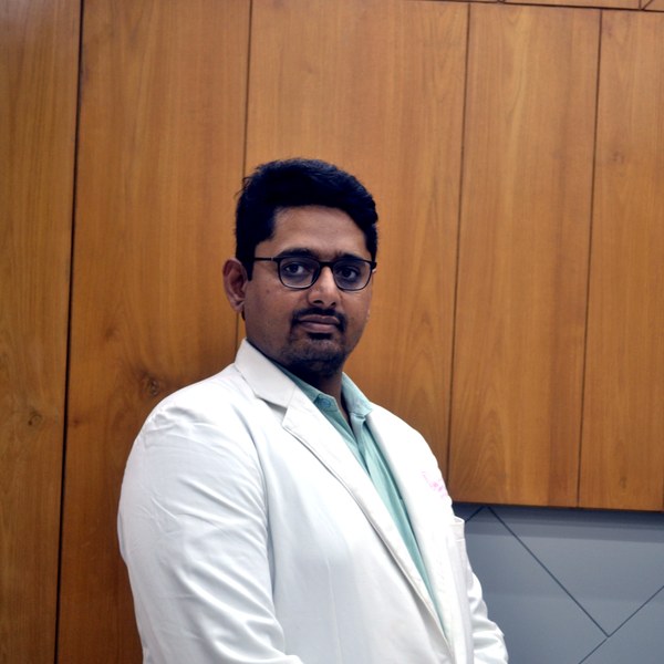
Dr Patel leads endometriosis diagnosis and surgery at Balaji Horizon with an evidence-based, ovarian-sparing philosophy aligned to ESHRE and ESGE — integrating pain, fertility and long-term disease control into a single plan, rather than treating the disease in isolation.
Imaging-led diagnosis, medical-first management, and precise surgery only when it is the right step — planned around your pain and fertility goals.
Dr. Priyadatt Patel
Senior Gynecologist · Advanced Laparoscopic Surgeon · IVF and Endometriosis Programme Lead
MS OBGyn · Pregnancy Care · Advanced Gynaecological Ultrasound · Fertility Preservation
ESHRE / ESGE / AAGL / ASRM guideline-aligned practice. 3D Karl Storz precision technique. Fertility-preservation-first philosophy. Evidence-based decisions, honest counselling, long-term outcomes orientation.
323, Satyamev Eminence, Beside Saptak Bungalows & AUDA Water Tank, Science City Road, Sola, Ahmedabad 380060
Open 24 hours (emergencies & labour) · OPD Mon–Sat 11:00–20:00 · +91 97234 31544
Naranpura, Ahmedabad
Mon–Sat 08:30–10:30 · +91 70460 02566