
Diagnostic Laparoscopy — When It Is Still Needed
Modern endometriosis practice has moved away from routine diagnostic laparoscopy. Expert imaging diagnoses most disease without surgery. Laparoscopy is now reserved for specific indications where it changes management — combined diagnosis-treatment, suspected disease with negative imaging in symptomatic patients, or specific histological questions.
1. Why diagnostic laparoscopy alone is no longer routine
Twenty years ago, laparoscopy was the only way to confirm endometriosis. Today, expert transvaginal ultrasound (IDEA protocol) and selective MRI diagnose most disease non-invasively. Routine diagnostic laparoscopy alone — without therapeutic intent — is no longer the standard of care per ESHRE 2022 and other current guidelines.
2. When laparoscopy is indicated
Symptomatic patient with normal imaging where suspicion remains high — typically combined with operative intervention in the same session if disease is found. Pre-IVF assessment in selected cases where pelvic adhesions or hydrosalpinx are suspected. Acute presentations (suspected endometrioma rupture, acute pelvic pain not yielding to other investigation). Histological confirmation when imaging is equivocal and impacts management.
3. The combined diagnostic-therapeutic approach
When laparoscopy is performed, it almost always combines diagnosis with treatment. Disease found is excised in the same session. This avoids repeated anaesthetic exposure. Pre-operative imaging (ultrasound, MRI) guides expectations and consent. The surgical team is prepared for therapeutic intervention.
4. Patient consent for combined procedures
Consent obtained for both diagnostic and treatment components. Patient briefed on imaging findings, expected disease pattern, and what therapeutic actions may be taken. Authorisation for excision, adhesiolysis, ovarian cystectomy, possible bowel/ureteric work where suspected. No surprise interventions.
5. Procedure technique
Under general anaesthesia. 3–5 small abdominal incisions. CO2 pneumoperitoneum. Diagnostic survey of entire pelvis and abdomen — peritoneum systematically inspected, ovaries assessed, pouch of Douglas examined, appendix and bowel surveyed. Findings photo-documented. Disease excised where appropriate per planned operative consent.
6. Findings and documentation
Visual identification of lesions (vesicular, red flame, black powder-burn, white scar). Mapping of disease per rASRM and #Enzian classifications. Biopsy of lesions for histological confirmation. Photo-documentation of pre and post-excision appearance. Operative findings shared with patient post-operatively with images.
7. Recovery from diagnostic laparoscopy
Day-surgery procedure in most cases. Discharge same day or next morning. Light activity 1–2 days. Office work 1–2 weeks for simple cases, longer if therapeutic excision was performed. Post-operative review at 2 weeks. Most patients recover quickly.
8. Negative laparoscopy in symptomatic patients
If laparoscopy is negative in a patient with significant symptoms, additional considerations: pelvic floor dysfunction (often coexists), bladder pain syndrome, IBS-overlap, neuropathic pain pathways with central sensitisation, gynaecological non-endometriosis causes. Negative laparoscopy redirects rather than ends the diagnostic journey.
When diagnostic laparoscopy is still needed
| Situation | Why |
|---|---|
| Negative imaging but high suspicion | Superficial disease is invisible on scans |
| Imaging–symptom mismatch | Clarify before major surgery |
| Planned therapeutic surgery | Diagnose and treat in one sitting |
| Unexplained infertility | Assess tubes and peritoneum |
Aligned with current international evidence, not habit.
Frequently Asked Questions
Do I need diagnostic laparoscopy?
What does laparoscopy show that imaging does not?
If I have laparoscopy, will I have treatment too?
What is the recovery time?
What are the risks?
Will laparoscopy leave scars?
What if laparoscopy is negative?
How is staging done at laparoscopy?
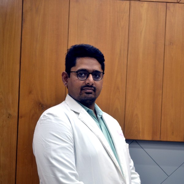
Dr Patel leads endometriosis diagnosis and surgery at Balaji Horizon with an evidence-based, ovarian-sparing philosophy aligned to ESHRE and ESGE — integrating pain, fertility and long-term disease control into a single plan, rather than treating the disease in isolation.
Imaging-led diagnosis, medical-first management, and precise surgery only when it is the right step — planned around your pain and fertility goals.
Dr. Priyadatt Patel
Senior Gynecologist · Advanced Laparoscopic Surgeon · IVF and Endometriosis Programme Lead
MS OBGyn · Pregnancy Care · Advanced Gynaecological Ultrasound · Fertility Preservation
ESHRE / ESGE / AAGL / ASRM guideline-aligned practice. 3D Karl Storz precision technique. Fertility-preservation-first philosophy. Evidence-based decisions, honest counselling, long-term outcomes orientation.
323, Satyamev Eminence, Beside Saptak Bungalows & AUDA Water Tank, Science City Road, Sola, Ahmedabad 380060
Open 24 hours (emergencies & labour) · OPD Mon–Sat 11:00–20:00 · +91 97234 31544
Naranpura, Ahmedabad
Mon–Sat 08:30–10:30 · +91 70460 02566