

Female Fertility Evaluation — The Comprehensive Workup
In short — A complete fertility workup covers ovarian reserve, the uterine cavity and tubes, ovulation and thyroid function, and a semen analysis for the partner. The purpose is to identify what is treatable before treatment is chosen, so that IVF is a considered step rather than a default.
A complete female fertility evaluation establishes ovarian reserve, ovulatory function, uterine cavity and tubal status, and identifies modifiable factors. This page describes the structured workup performed before any IVF or assisted conception decision — aligned with ESHRE 2023 and ASRM 2024 guidance.
1. Detailed clinical history
Menstrual history (cycle length, regularity, pain, flow), obstetric history, sexual and contraception history, surgical history (especially pelvic surgery), family history of premature ovarian insufficiency or endometriosis, medical conditions (thyroid, diabetes, PCOS, autoimmune), lifestyle (weight, smoking, alcohol, exercise, stress, sleep), and partner factors. The history alone often reveals 60–70 percent of the diagnostic answer.
2. AMH — anti-Müllerian hormone
AMH reflects the size of the remaining follicle pool. Random blood test, no cycle-day requirement. Interpretation: above 3.0 ng/ml suggests potential PCOS or high responder; 1.0–3.0 ng/ml normal range; 0.5–1.0 ng/ml diminished ovarian reserve; below 0.5 ng/ml severely diminished. AMH predicts IVF response (number of eggs retrieved) better than it predicts pregnancy chance.
3. Antral follicle count (AFC)
Transvaginal ultrasound counting follicles 2–10 mm in both ovaries, performed in early follicular phase (cycle day 2–5). Total AFC of 12–25 is normal range, above 25 suggests PCOS or high responder, below 7 suggests diminished reserve. AFC and AMH together provide the most accurate ovarian reserve picture.
4. Day 2–3 hormone profile
FSH below 10 mIU/ml is reassuring; above 10 suggests diminished reserve. Estradiol above 60 pg/ml early in cycle suggests poor response despite normal-appearing FSH. LH evaluated for PCOS pattern. TSH below 2.5 mIU/L is optimal for fertility (treat subclinical hypothyroidism). Prolactin checked once to exclude hyperprolactinaemia.
5. Uterine cavity assessment
Transvaginal ultrasound assesses general cavity, myometrial thickness, fibroids, polyps. Saline-infusion sonography (SIS) — gold standard for cavity assessment, identifies sub-mucous fibroids, polyps, adhesions, congenital anomalies. Hysteroscopy reserved for diagnostic confirmation, surgical correction, or chronic endometritis biopsy in selected cases.
6. Tubal patency assessment
Hysterosalpingography (HSG) — radiographic test of tubal patency, also assesses cavity. HyCoSy (saline-infusion sonography with foam contrast) — ultrasound-based alternative, avoids radiation. Laparoscopy with dye (chromopertubation) — gold standard, reserved for cases needing operative intervention. Hydrosalpinx identified must be addressed before IVF (clipping or salpingectomy).
7. Expert pelvic ultrasound for endometriosis
ISUOG IDEA consensus protocol — systematic transvaginal ultrasound mapping for endometriosis. Identifies endometriomas, deep infiltrating disease, ovarian mobility (kissing-ovaries sign), pouch of Douglas obliteration. Critical for fertility-seeking patients because endometriosis affects ovarian reserve, embryo quality and implantation.
8. Additional tests in selected cases
Chronic endometritis (CD138 immunohistochemistry on hysteroscopic biopsy) for recurrent failure. Karyotype both partners in recurrent loss. Antiphospholipid antibodies in recurrent loss or implantation failure. Thrombophilia panel in personal/family history of thrombosis. Sperm DNA fragmentation when standard semen analysis is normal but unexplained subfertility exists. Genetic carrier screening for selected ancestries.
The comprehensive workup
| Domain | Tests |
|---|---|
| Ovarian reserve | AMH, antral follicle count |
| Uterus | Scan, cavity assessment |
| Tubes | Where relevant |
| Male | Semen analysis |
Our IVF practice follows international reproductive-medicine standards.
Frequently Asked Questions
What does the female fertility workup involve?
Is AMH a measure of egg quality?
What if my AMH is low?
Do I need HSG or HyCoSy?
How long does the evaluation take?
Is laparoscopy needed for diagnosis?
What about recurrent miscarriage workup?
How does evaluation differ for unexplained infertility?
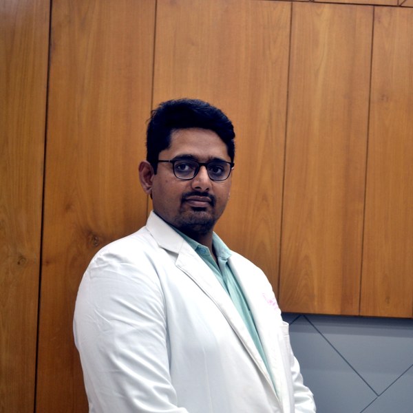
Dr Patel leads fertility care at Balaji Horizon, integrating reproductive surgery and IVF into a single plan — ethical, evidence-based and individualised, with realistic expectations and no overpromising of success.
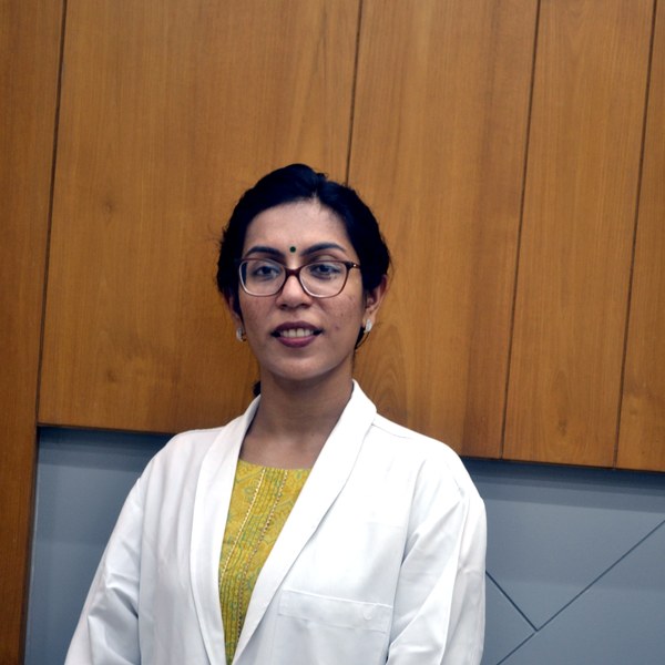
Individualised IVF and fertility planning with honest, evidence-based counselling — and realistic expectations from the very first consultation.
Dr. Priyadatt Patel
Senior Gynecologist · Advanced Laparoscopic Surgeon · IVF and Endometriosis Programme Lead
MS OBGyn · Pregnancy Care · Advanced Gynaecological Ultrasound · Fertility Preservation
ESHRE / ESGE / AAGL / ASRM guideline-aligned practice. 3D Karl Storz precision technique. Fertility-preservation-first philosophy. Evidence-based decisions, honest counselling, long-term outcomes orientation.
Science City Road, Ahmedabad 380060
Mon–Sat 11:00–20:00 · +91 97234 31544
Naranpura, Ahmedabad
Mon–Sat 08:30–10:30 · +91 70460 02566